

Pages
Rivelazione e caratterizzazione di inquinanti tossici in acqua di mare ed animali marini
PROGETTI RETROSPETTIVI COERENTI CON LA MISURA 3.5 DEL FEP CAMPANIA 2007 – 2013 (ex D.D. del 21.12.2015, n° 854)
Responsabile Scientifico: Giovanna Romano
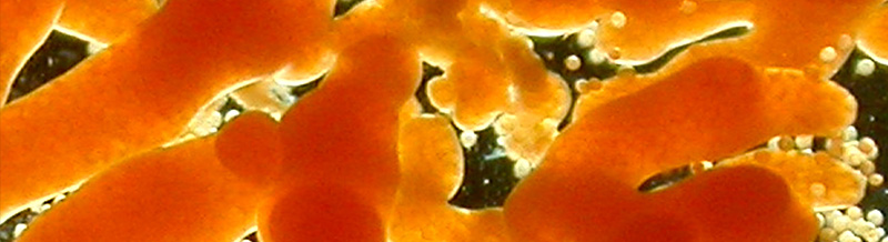
Il progetto Rivelazione e caratterizzazione di inquinanti tossici in acqua di mare ed animali marini ha affrontato tematiche relative all’inquinamento ambientale con particolare riguardo all’inquinamento delle acque costiere marine da metalli pesanti ed idrocarburi policiclici aromatici, per l’impatto enorme che la presenza di queste sostanze tossiche può avere sulla salute dell’uomo, sia mediante contatto diretto sia attraverso il consumo di pesce e molluschi per uso alimentare.
Il progetto è stato finanziato nell'ambito della MISURA 3.5 DEL FEP CAMPANIA 2007 – 2013, (ex D.D. del 21.12.2015, n° 854).
Gli Istituti partner del progetto hanno svolto attività di ricerca focalizzate sulla messa a punto di metodi biochimici, biofisici e biologici per la determinazione sia del mercurio che di alcuni IPA, in acqua di mare o in matrici che ne mimano la composizione.
Le azioni previste dal Progetto hanno mirato in particolare alla caratterizzazione delle proprietà tossicologiche del mercurio in ricci di mare e copepodi nonché alla raccolta di dati analitici di concentrazione di mercurio e derivati di mercurio in molluschi cefalopodi per la caratterizzazione dei livelli di bio-accumulo di metalli in traccia e di sostanze derivate dall’attività umana allo scopo di individuare le eventuali possibili ricadute sul benessere animale e non da ultimo sulla “biosicurezza”.
Il nostro ruolo: La Stazione Zoologica ha coordinato il progetto ed è stata responsabile dell’obiettivo 3 (Valutazione della tossicità del mercurio e altri metalli pesanti su organismi marini di rilevanza ecologica) e dell’obiettivo 5 (Raccolta di dati analitici di concentrazione di mercurio e derivati di mercurio per la caratterizzazione dei livelli di bio-accumulo di metalli in cefalopodi).
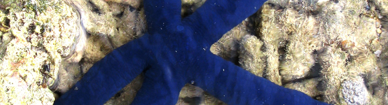
Gli obiettivi principali sono stati la valutazione degli effetti di mercurio e altri metalli pesanti su organismi marini che rivestono un ruolo ecologico di grande rilevanza per nella colonna d’acqua (copepodi) e sui fondali, a diretto contatto dei sedimenti (riccio di mare). In considerazione del fondamentale ruolo dei copepodi e dei ricci nell’ambiente marino, abbiamo, condotto uno con l’obiettivo di ampliare il numero di ‘stress genes’ da impiegare come biomarker negli studi ecotossicologici. Sono stati valutati inoltre gli effetti della concentrazione di mercurio in diversi tessuti di due specie di cefalopodi (polpi, Octopus vulgaris; seppie, Sepia officinalis) ottenuti dal pescato locale. Un obiettivo collaterale di questo studio è stato quello di poter stabilire valori stimati di concentrazione di mercurio e suoi metaboliti in tessuti di cefalopodi in Tirreno (Golfo di Napoli) potendo così contribuire alla creazione di un network internazionale che possa contribuire ad uno studio “epidemiologico” sull’impatto delle concentrazioni di metalli pesanti e in traccia, tra cui il mercurio, su alcune specie chiave del pescato mondiale, quali proprio polpi e calamari.
Partners: Istituto di Biostrutture e Bioimmagini del CNR (CNR-IBB), Dipartimento di Biochimica, Biofisica e Patologia Generale (SUN-DBBPG), Dipartimento di Scienze e Tecnologie Ambientali, Biologiche e Farmaceutiche della Seconda Università degli Studi Napoli (SUN-DISTABIF), Stazione Zoologica Anton Dohrn (SZN) e Dipartimento di Fisica dell’Università Federico II di Napoli (UNINA-DIPFIS).
Publications
2020
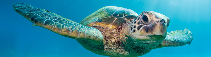
Marchiori E, Dotto G, Tessarin C, Santoro M, Affuso A, Tarricone L, Di Renzo L, Freggi D, Spoto V, Marcer F (2020) A pilot study on molecular diagnosis of Hapalotrema mistroides (Digenea: Spirorchiidae) infection in blood samples of live loggerhead turtles Caretta caretta. BMC Veterinary Research 16: 16. https://doi.org/10.1186/s12917-020-2232-y
2019
Arcangeli A, Maffucci F, Atzori F, Azzolin M, Campana I, Carosso L, Crosti R, Frau F, David L, Di-Méglio N, Roul M, Gregorietti M, Mazzucato V, Pellegrino G, Giacoletti A, Paraboschi M, Zampollo A, de Lucia GA, Hochscheid S (2019) Turtles on the trash track: loggerhead turtles exposed to floating plastic in the Mediterranean Sea. Endangered Species Research 40: 107-121. DOI: https://doi.org/10.3354/esr00980
Hochscheid S., M. Aksissou, T. Arapis, M. Benabdi, L. Boura, A. Broderick, L. Cardona, C. Carreras, F. Claro, A. Demetropoulos, W. J. Fuller, I. Jribi, Y. Kaska, Y. Levy, F. Maffucci, D. Margaritoulis, C. Mifsud, A. Panagopoulou, J. Sacchi, J. Tomás, O. Türkozan, A. Rees (2019) Sea Turtles of the Mediterranean Sea. Special Feature – State of the World’s Sea Turtles SWOT Report XIV, Oceanic Society, Ross, CA, USA, pp. 20-29. https://www.seaturtlestatus.org/swot-report-vol-14
Pace A, Dipineto L, Fioretti A, Hochscheid S (2019) Loggerhead sea turtles as sentinels in the western Mediterranean: antibiotic resistance and environment-related modifications of Gram-negative bacteria. Marine Pollution Bulletin 149: 110575. https://doi.org/10.1016/j.marpolbul.2019.110575
Pace A, Rinaldi L, Ianniello D, Borrelli L, Cringoli G, Fioretti A, Hochscheid S, Dipineto L (2019) Gastrointestinal investigation of parasites and Enterobacteriaceae in loggerhead sea turtles from Italian coasts. BMC Veterinary Research 15: 370. https://doi.org/10.1186/s12917-019-2113-4
Riquet F, Liautard-Haag C, Woodall L, Bouza C, Louisy P, Hamer B, Otero-Ferrer F, Aublanc P, Béduneau V, Briard O, El Ayari T, Hochscheid S, Belkhir K, Arnaud-Haond S, Gagnaire P-A, Bierne N (2019) Parallel pattern of differentiation at a genomic island shared between clinal and mosaic hybrid zones in a complex of cryptic seahorse lineages. Evolution 73: 817-835. https://doi.org/10.1111/evo.13696
Publications
2020
Biasetti P, Florio D, Gili C, & de Mori B (2020) The Ethical Assessment of Touch Pools in Aquariums by Means of the Ethical Matrix. Journal of Agricultural and Environmental Ethics 33(2):337-353. https://doi.org/10.1007/s10806-020-09823-2
Camiñas, J.A.; Kaska, Y.; Hochscheid, S.; Casale P.; Panagopoulou, A.; Báez, J.C.; Otero, M. M.; Numa, C., Alcázar, E. (2020). Conservation of marine turtles in the Mediterranean sea [brochure]. IUCN, Malaga, Spain. https://www.iucn.org/sites/dev/files/content/documents/2020/conservation_of_mediterranean_turtles_in_the_mediterranean_sea.pdf
Casale P., Hochscheid S., Kaska Y., Panagopoulou A. (Eds.) (2020). Sea Turtles in the Mediterranean Region: MTSG Annual Regional Report 2020. Report of the IUCN-SSC Marine Turtle Specialist Group, 2020.
Centelleghe, C., Carraro, L., Gonzalvo, J., Rosso, M., Esposti, E., Gili, C., Bonato, M., Pedrotti, D., Cardazzo, B., Povinelli, M., & Mazzariol, S. (2020). The use of Unmanned Aerial Vehicles (UAVs) to sample the blow microbiome of small cetaceans. PloS one, 15(7), e0235537. https://doi.org/10.1371/journal.pone.0235537
Chimienti M., M.F. Blasi, S. Hochscheid (2020) Movement patterns of large juvenile loggerhead turtles in the Mediterranean Sea: ontogenetic space use in a small ocean basin. Ecology and Evolution 10(14), 6978- 6992. DOI:10.1002/ece3.6370
Krahl A., A. Lipphaus, P. Martin Sander, F. Maffuci, S. Hochscheid, U. Witzel (2020) Humerus osteology, myology, and finite element structure analysis of Cheloniidae. The Anatomical Record 303, 2177–2191. DOI:10.1002/ar.24311
Leonetti FL, Sperone E, Travaglini A, Mojetta AR, Signore M, Psomadakis PN, Dinkel TM, & Bottaro M (2020) Filling the Gap and Improving Conservation: How IUCN Red Lists and Historical Scientific Data Can Shed More Light on Threatened Sharks in the Italian Seas. Diversity 12(10):389. https://doi.org/10.3390/d12100389
Maffucci F., A. Pace, A. Affuso, M. Ciampa, G. Treglia, A. Pignalosa, S. Hochscheid (2020) Carapace scute pattern anomalies in the loggerhead turtle: are they indicative of hatchling’s survival probability? Journal of Zoology, 310(4), 315-322. https://doi.org/10.1111/jzo.12754
Marchiori E, Dotto G, Tessarin C, Santoro M, Affuso A, Tarricone L, Di Renzo L, Freggi D, Spoto V, Marcer F (2020) A pilot study on molecular diagnosis of Hapalotrema mistroides (Digenea: Spirorchiidae) infection in blood samples of live loggerhead turtles Caretta caretta. BMC Veterinary Research 16: 16. https://doi.org/10.1186/s12917-020-2232-y
Mennonna, G., B. Lamagna, A. Affuso, A. Greco, F. Micieli, D. Costanza, S. Hochscheid, L. Meomartino (2020) Normal ultrasonographic features of loggerhead (Caretta caretta) eyes. Natura Croatica, 29, Suppl. 1., 3-10. DOI: 10.20302/NC.2020.29.18
2019
Arcangeli A, Maffucci F, Atzori F, Azzolin M, Campana I, Carosso L, Crosti R, Frau F, David L, Di-Méglio N, Roul M, Gregorietti M, Mazzucato V, Pellegrino G, Giacoletti A, Paraboschi M, Zampollo A, de Lucia GA, Hochscheid S (2019) Turtles on the trash track: loggerhead turtles exposed to floating plastic in the Mediterranean Sea. Endangered Species Research 40: 107-121. DOI: https://doi.org/10.3354/esr00980
Hochscheid S., M. Aksissou, T. Arapis, M. Benabdi, L. Boura, A. Broderick, L. Cardona, C. Carreras, F. Claro, A. Demetropoulos, W. J. Fuller, I. Jribi, Y. Kaska, Y. Levy, F. Maffucci, D. Margaritoulis, C. Mifsud, A. Panagopoulou, J. Sacchi, J. Tomás, O. Türkozan, A. Rees (2019) Sea Turtles of the Mediterranean Sea. Special Feature – State of the World’s Sea Turtles SWOT Report XIV, Oceanic Society, Ross, CA, USA, pp. 20-29. https://www.seaturtlestatus.org/swot-report-vol-14
Pace A, Dipineto L, Fioretti A, Hochscheid S (2019) Loggerhead sea turtles as sentinels in the western Mediterranean: antibiotic resistance and environment-related modifications of Gram-negative bacteria. Marine Pollution Bulletin 149: 110575. https://doi.org/10.1016/j.marpolbul.2019.110575
Pace A, Rinaldi L, Ianniello D, Borrelli L, Cringoli G, Fioretti A, Hochscheid S, Dipineto L (2019) Gastrointestinal investigation of parasites and Enterobacteriaceae in loggerhead sea turtles from Italian coasts. BMC Veterinary Research 15: 370. https://doi.org/10.1186/s12917-019-2113-4
Riquet F, Liautard-Haag C, Woodall L, Bouza C, Louisy P, Hamer B, Otero-Ferrer F, Aublanc P, Béduneau V, Briard O, El Ayari T, Hochscheid S, Belkhir K, Arnaud-Haond S, Gagnaire P-A, Bierne N (2019) Parallel pattern of differentiation at a genomic island shared between clinal and mosaic hybrid zones in a complex of cryptic seahorse lineages. Evolution 73: 817-835. https://doi.org/10.1111/evo.13696
Third Mission Area
Naples Seat
Massimo Cavaliere (Coordinatore dell’Area a.i.)
Communication, Training, Technology Transfer and Dissemination Section
Higher Education
Gabriella Grossi, Coordinator
Margherita Groeben
Francesca Motti
Grant Innovation Office (GIO)
Naples
Giorgio Carpino, Coordinator
Danilo Cavaliere
Alberto Corona
Roberto Firmamento
Martina Genovese
Valerio Mattera
Ornella Papaluca
Paola Punzo
Giosuè Zurzolo
Institutional communication
NO affilations
Facilities Open to the Public Section
Naples Seat - DaDoM
General Administration Area
Sede Roma
• Francesca Di Carlo (Rome Coordinator)
Sede Napoli
• Gaetano Aloe
• Valeria Contino
• Nicola Manco
• Chiara Svampa
General Services
Administrative Services
Technical Services
Central Administration
General Administration Area - Coordinator ad interim: Director General
Third Mission Area - Coordinator ad interim: Director General
Fourteenth Ischia Summer School on the History of the Life Sciences
Fourteenth Ischia Summer School - 27 June – 3 July 2015
Geographies of Life
Introduction to the theme
Life’s diversity is today an integral part of the various climates and locales our planet has to offer. Herodotus wrote of the stations of the earth’s life forms, and since Aristotle the sea has also attracted naturalists as a source of wonders that confound land-based classifications. Yet understandings of the spatial distribution of life have changed radically over time. In the ancient world, land and sea formed separate spheres in a structured cosmos of “natural places,” each of which possessed its properly adapted inhabitants. For Aristotle, seals were “monsters,” because they show all the main features of land animals, but live in the wrong place. Living beings could be in the right place or out of place, they could inhabit temperate and marginal (hot or cold) zones, but the patterns were not understood in terms of geographic distribution on a grid of latitude and longitude.
Early modern voyages of exploration added this geographical dimension. Sea and land collapsed into one “terraqueous globe,” and naturalists began to realize that identical climes could harbour very different fauna and flora. At the same time, the concept of species acquired temporal and spatial dimensionality, with species now understood as physical and physiological systems in their own right, rather than forms that matter could take on. Only in the nineteenth century, however, did the spatial distribution of organisms become the subject of a dedicated field of research, biogeography. Alexander von Humboldt’s attempt to derive quantitative biogeographic “laws” led to the realization that the distribution of species did not simply follow the physical environment as it varied with latitude, altitude, and geological conditions, but was the contingent result of migrations, displacements, and hybridisations. Evolutionism, that is, depended not only on the discovery of “deep time” (itself a spatial metaphor), but also on the temporalisation and dynamisation of spatial relations. The consolidation of nation states, as well as colonial and imperial projects, was the political correlate of this development, which was equally visible in the human sciences, with medical topographies feeding into epidemiology, and racial typologies into anthropology and demography.
From the late nineteenth century, when the sea also acquired layers of depth and a detailed topography, an international network of field stations were dedicated, for example, to marine biological and high-altitude research. These institutions facilitated in situ investigations of living organisms and the study of human bodies under extreme conditions. Colonial and imperial surveys, the promotion of agriculture and fisheries by nation states, epidemiology and population genetics, the integration of meteorology and hydrology into climatology, and finally, the use of radioactive isotopes and satellite data in tracking life on a global scale, have turned geographic space into an integral and essential component of contemporary understandings of life on earth. Thus, if the nineteenth century saw the dynamisation of geographic space, the twentieth century saw its experimentalisation, the turning of landscapes into ‘labscapes’, as Robert Kohler called them.
Historians have studied the geographic dimension of the life sciences from a diversity of perspectives, though usually with a focus on particular fields: natural history in the context of exploration and empire, biogeography, oceanography, ecology, epidemiology, demography and medical geography. This summer school adds perspectives from the spatial turn in the history of science, medicine and technology, including studies of transregional and global exchange networks, which have often taken inspiration from imperial studies, oceanic histories, and world history. It also takes account of spatially organized inscription devices, including the lists, catalogues, maps, statistical records, and databases that can synoptically present data gathered from various places.
It was timely to explore the changing relationship between humans and the spatially organized environment also because, confronted by problems of disease control, food security, conservation biology, and climate change, the biosciences themselves increasingly study life as a complex, spatially distributed phenomenon, be it on the micro-scale of biofilms and gut floras, or the macro-scale of the biosphere. This may represent a reawakening after a period when molecular biology dominated, or developments of research programmes that were always alternatives to the molecular paradigm, or the opening up of new spaces for research by the very molecularization of life. At the same time, human geographers had turned their attention to the life sciences as a phenomenon to be addressed with their own tools. Though such concepts as Friedrich Ratzel’s Lebensraum have a long (and problematic) history, geographers had recently begun to study the production of biological knowledge in its own right. Often taking spatial metaphors in the life sciences as a starting point – “boundary,” with its prominent place in immunology, is a telling example – they were exploring the co-production of spatial relations through interactions between humans, both experts and laypeople, and other organisms. The summer school on “Geographies of Life” thus addressed a subject of urgent relevance to the evolving relations of humans with our natural and social environments, and added historical depth to attempts to understand the roles of the life sciences in changing those relations.
1st International Summer School on Marine Ecotoxicology (SSME1)
The first International Summer School on Marine ecotoxicology, SSME1, is organized by the Stazione Zoologica Anton Dohrn under the patronage of Ordine Nazionale dei Biologi and will take place in July 2019 from the 1st to the 5th.
SSME1 will provide an advanced training on different classes of traditional and emerging marine pollutants and how biological systems respond to and are affected by these contaminants at all biological levels.
The participants will focus on:
- Marine chemical stressors: metals, antifoulants, nanomaterials, plastics
- Climate changes: global warming, ocean acidification
- Marine natural toxins
- Marine model organisms in ecotoxicology
- Biomarkers and bioindicators in marine environmental monitoring
- Reprotoxicology
- Case studies
- Ecotoxicity tests
The program consists of lectures and practical sessions held by internationally recognized experts and researchers in the field. Practical session will include innovative and standardized toxicity tests for the assessment of ecotoxicological effects in different marine organisms.
This course will allow the participants to acquire methods and approaches for the detection and assessment of the impact of environmental stressors on marine biota.
SSME1 is open to 20 people with basic knowledge in ecotoxicology
The registration fee is 300,00€ (three hundreds/00), which includes documentation, coffee and lunches, but it does not include accommodation.
The Ordine Nazionale dei Biologi (ONB) supports SSME1 with funds to cover the fee for thirteen participants and provides a certificate worth ECM credits (continued medical education) to all participants.
Summer School “Protein Evolution”
Protein Evolution: from Environmental Adaptations to Biotechnological Applications
A Summer School on “Protein Evolution” will be held at the Stazione Zoologica Anton Dohrn in Naples on July 24 - 26. The Program will include lectures on theoretical aspects of molecular evolution, sequence data manipulation and alignment, protein structure, molecular modelling, and docking analysis. Lectures will be integrated with practical tutorials; and participants are encouraged to work on their own laptop. The number of participants will be limited to 22, and admission is on a first come, first served basis.
Publications
Copyright © 2015 by Stazione Zoologica Anton Dohrn Napoli - Italy
Villa Comunale, 80121 Napoli - Tel.+39 081 5833111 - C.F./P.IVA : IT 04894530635 - PEC: ufficio.protocollo(at)cert.szn.it