

News
The PhD project will be conducted in collaboration between Università degli Studi di Ferrara and Stazione Zoologica Anton Dohrn.
We invite applications for a PhD student to work with Dr. Salvatore D’Aniello (Stazione Zoologica Anton Dohrn of Napoli) and Prof.Cristiano Bertolucci (University of Ferrara) on “The circadian clock in the deep-sea fish”. The student will have the opportunity to spend 6 months in secondment in Nick Foulkes lab at the Karlsruhe Institute of Technology (Germany).
To apply, please follow this link and the guidelines therein.
For more information, please contact us at This email address is being protected from spambots. You need JavaScript enabled to view it. and This email address is being protected from spambots. You need JavaScript enabled to view it.
The application deadline is September 2nd, 2024, 12.00 CET
The PhD project will be conducted in collaboration between the Università Politecnica and the Stazione Zoologica Anton Dohrn.
We invite applications for a PhD student to work with Dr Carlo Cattano on “Past, present and future of Elasmobranchs in Italian waters: Trends in abundance, species Occurrence and Resource Use (TORU)”
To apply, please follow this link and the guidelines therein.
The application deadline is August 2nd, 2024, 01.00 P.M. CET
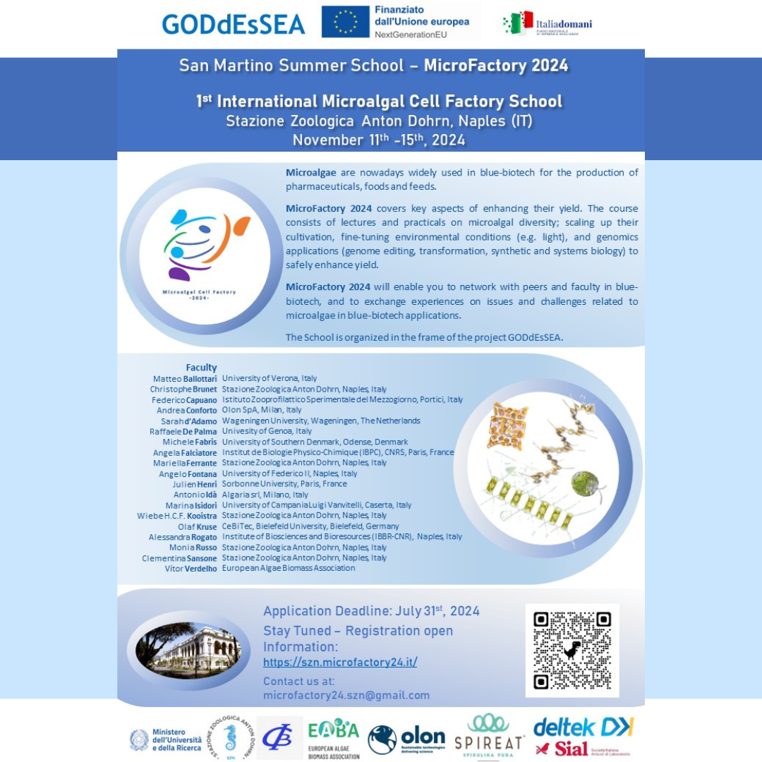
???? Dates: November 11th to 15th, 2024
???? Location: Stazione Zoologica Anton Dohrn, Naples, Italy
We are excited to announce the FIRST EDITION of the International Summer School on Microalgal Cell Factory, designed for Ph.D. students and young researchers. This event aims to build a lively scientific community tackling global challenges in using microalgae as biofactories of natural products for biotechnological applications.
Why Attend MicroFactory 2024?
Networking: Connect with peers and faculty in blue-biotech.
Knowledge Exchange: Share experiences on issues and challenges related to microalgae in blue-biotech applications.
Comprehensive Course: Includes lectures and practicals on microalgal diversity, scaling up cultivation, fine-tuning environmental conditions (e.g., light), and genomics applications (genome editing, transformation, synthetic and systems biology).
This will be a fantastic occasion to build a new and stimulating international network and to present your research activities.
Application Deadline: July 31th, 2024 Stay Tuned –
Registration open Information: https://szn.microfactory24.it/
Contact us at: This email address is being protected from spambots. You need JavaScript enabled to view it.
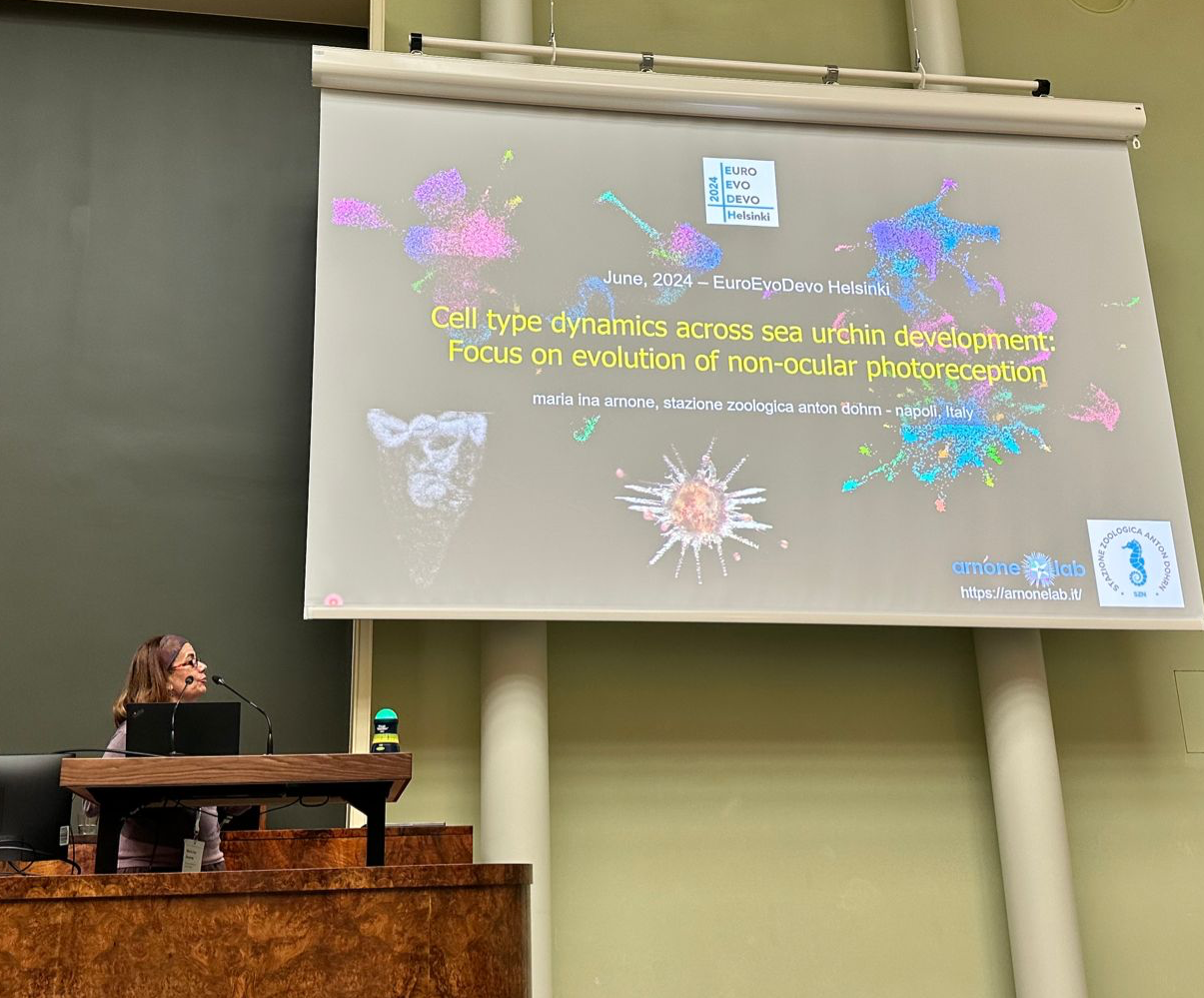
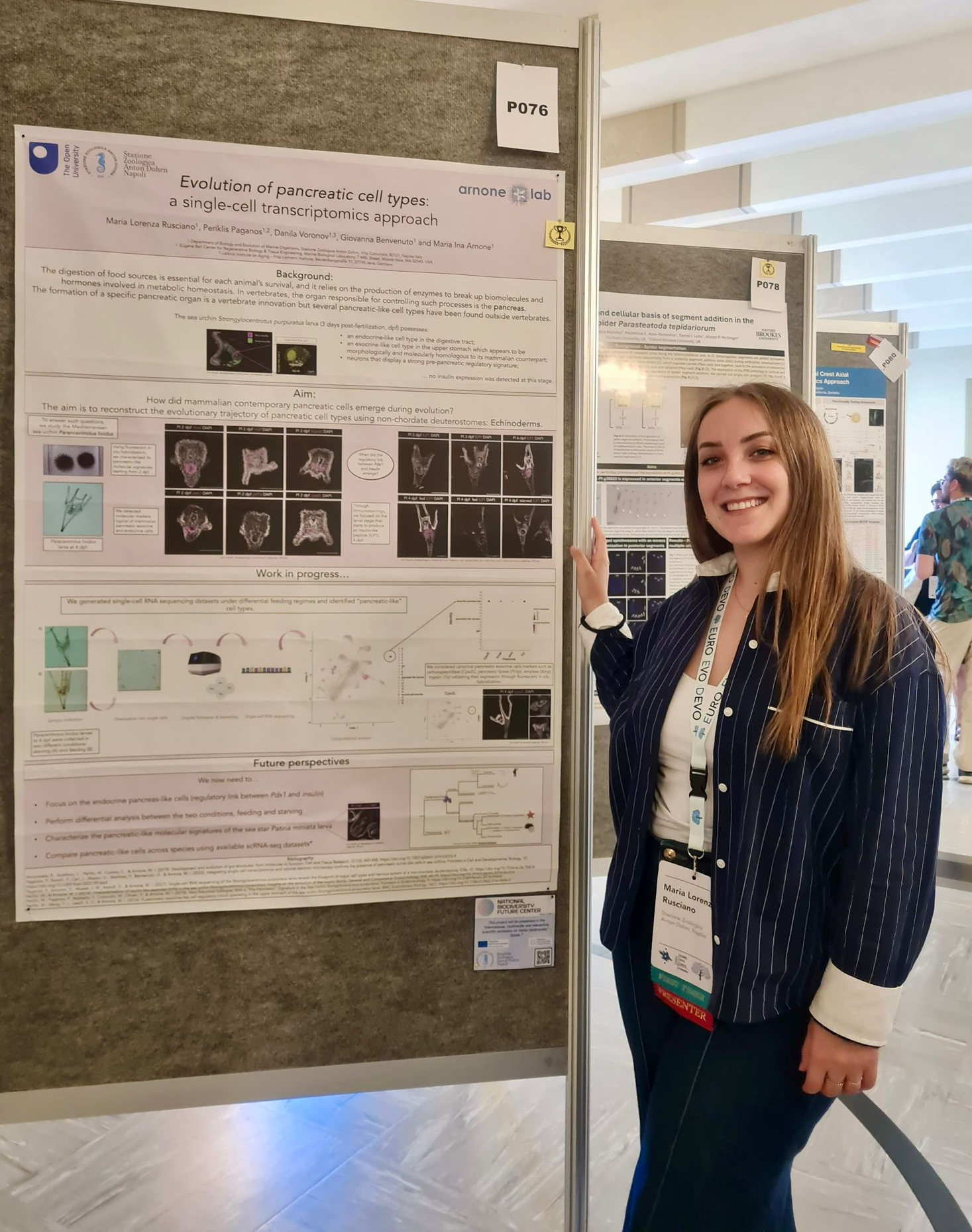
The 9th Conference of the European Society for Evolutionary Developmental Biology, EuroEvoDevo (https://www.helsinki.fi/en/conferences/euroevodevo-2024), has been held last week in the beautiful city of Helsinki, Finland. A diversity of fascinating questions, organisms and technologies were discussed at the meeting attended by more than 600 delegates from all over the world.
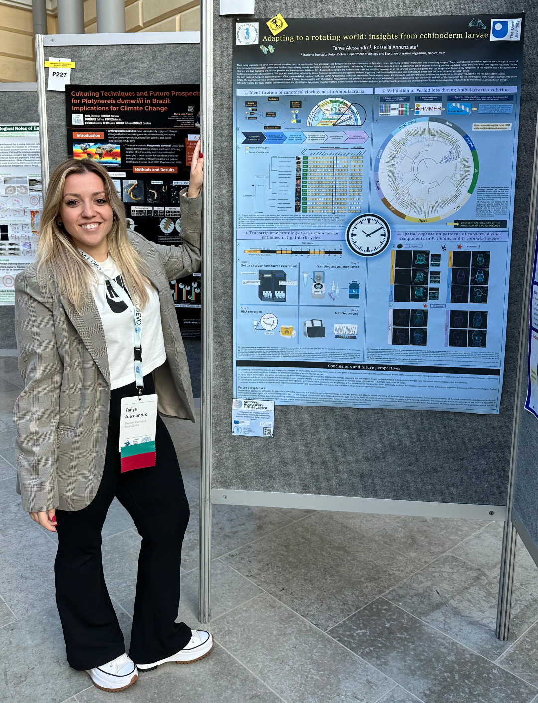
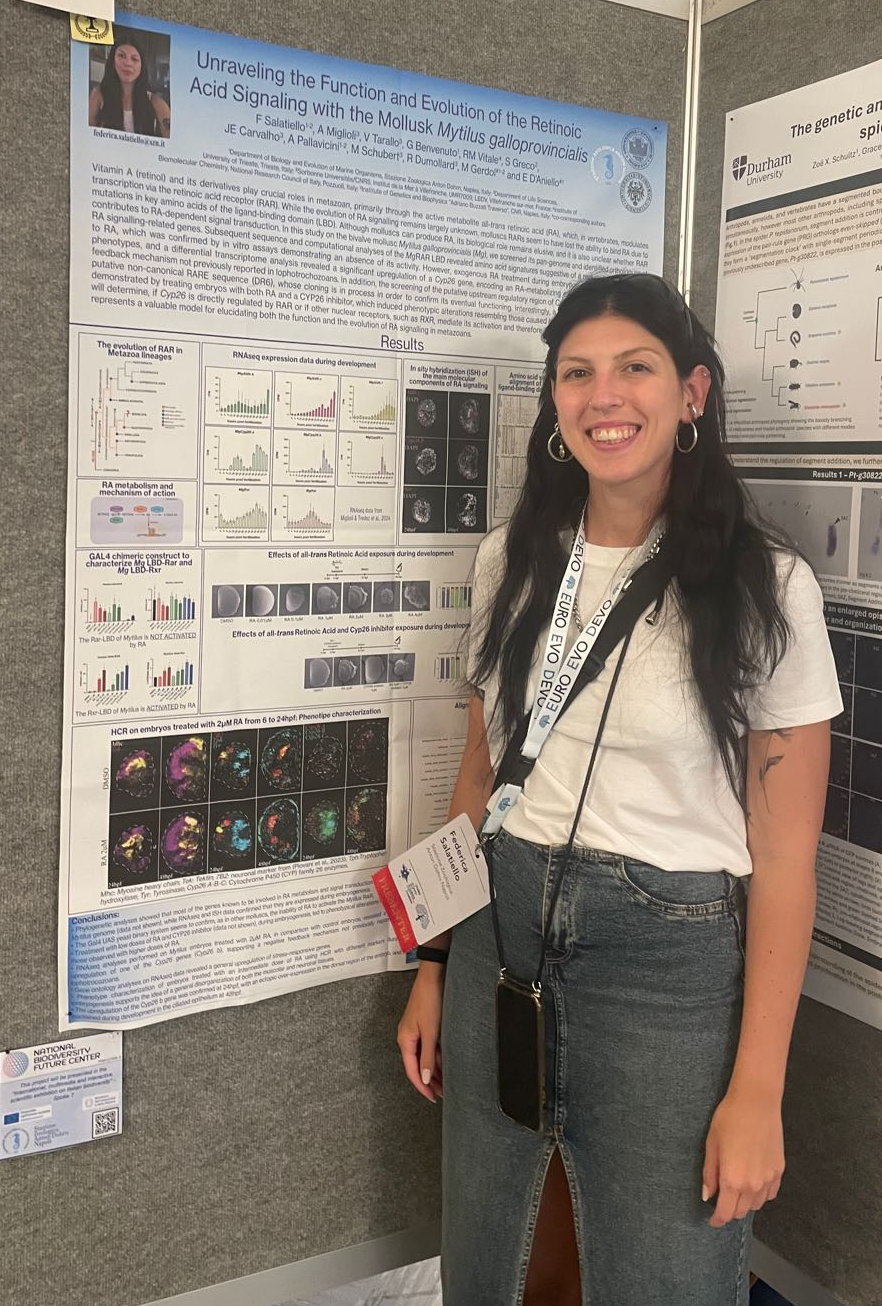
SZN was present with 12 colleagues and their contributions featured two exciting talks (Ina Arnone and Maria Cocurullo) and one poster (Lorenza Rusciano) on sea urchin development, an award-winning poster on the evolutionary role of retinoic acid during mussel embryogenesis (Federica Salatiello), and yet another poster on retinoic acid and its function in cardio-pharyngeal specification during-tunicates embryogenesis (Enrico D’Aniello). The Golgi structural changes in development and differentiation (Giovanna Benvenuto), light sensing in Octopus (Giacinto De Vivo) and the circadian clock in echinoderm larvae (Tanya Alessandro) were also lively introduced at the conference through poster presentations. Even diatoms made it to the conference, with two well received oral presentations on diatom life cycles and sexual reproduction (Mariella Ferrante and Antonella Ruggiero), and a cool poster on the response to environmental stress (Alessandra Mussi)!
SZN (Salvatore D’Aniello) was also partner in the organization of the “Amphioxus Satellite Meeting” that has seen a growing interest in comparative studies of marine invertebrate deuterostomes.
Get in touch with the SZN Researchers if you are interested in learning more on these topics.
One of the main challenges of marine organisms that inhabit the extreme-cold Antarctic waters is to stay alive without freezing. An entirely Italian research team led by Prof. Cinzia Corinaldesi of the Polytechnic University of Marche, together with colleagues from the Stazione Zoologica Anton Dohrn (Dr. Emanuela Buschi, first author of the pubblication, and the researchers Micheal Tangherlini and Sergio Stefanni), from Universities of Bologna, Ferrara and Salento, reveals, for the first time, that three endemic species of marine polychaete worms are able to resist extreme cold thanks to the production of antifreeze proteins produced by symbiotic bacteria living inside them. Researchers have discovered particular species of bacteria that are hosted, in great abundance, in the tissues of some Antarctic polychaetes. These bacteria produce antifreeze proteins, i.e. special proteins that lower the freezing point of internal liquids and make them more fluid, making "blood" circulation more efficient and better oxygenation of the animal, allowing it to adapt at low temperatures.
The study was published in Science Advances under the title “Resistance to freezing conditions of endemic Antarctic polychaetes is enhanced by cryoprotective proteins produced by their microbiome.”
1. Antarctic worms. Credits by Michael Tangherlini (SZN)
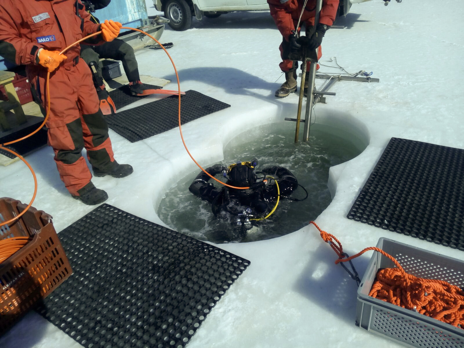
2. Sampling activity in Antarctica. Credits by Marco Lo Martire (UNIVPM)
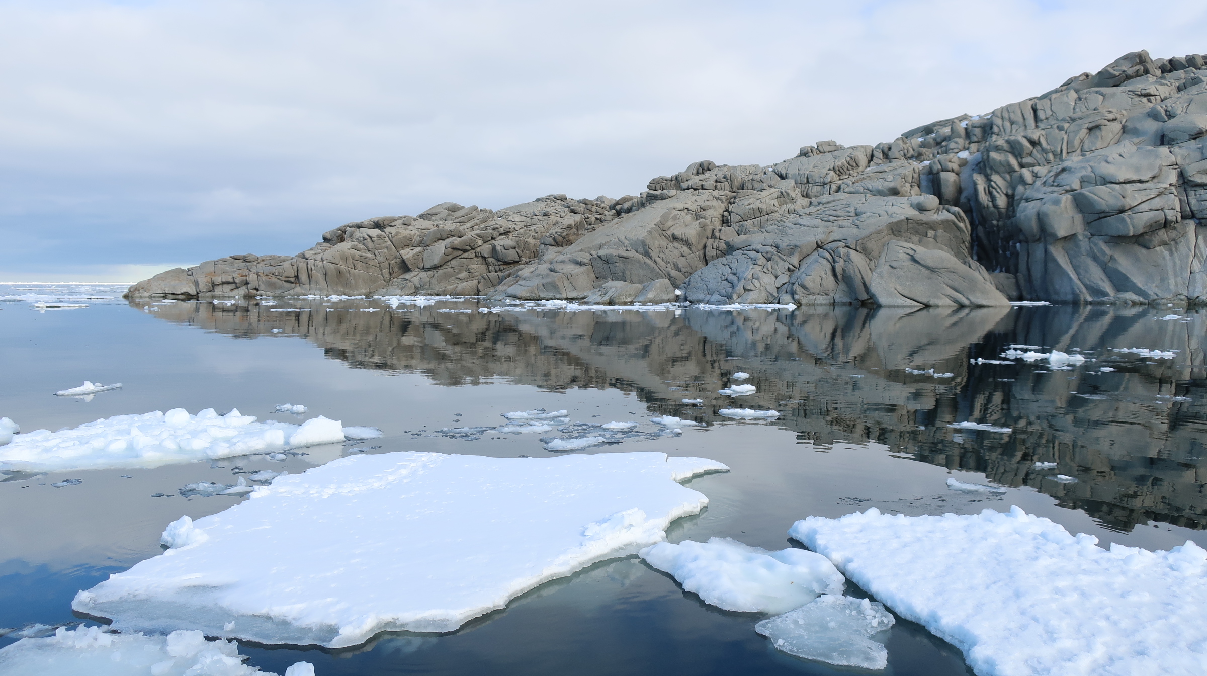
3. Antarctic landscape (Ross Sea). Credits by Marco Lo Martire (UNIVPM)
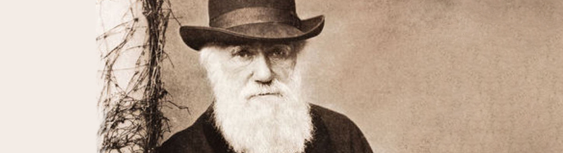
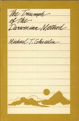
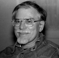 On June 14, 2024, the biologist, philosopher and historian of science Michael T. Ghiselin, member of the California Academy of Sciences, passed away forever. His specialty and passion were nudibranchs and he was known for his criticism of the falsification of the history of Lamarckism in biology textbooks. The Stazione Zoologica (SZN) has lost another member of its family. In September 1968 Ghiselin came to the SZN for two months, taking advantage of the AIBS (American Institute of Biological Sciences) table to - we read on his file card - "use the primary sources existing in the library [the archive was not yet there] to historical studies on the influence of the theory of evolution on 19th century embryology”. The following year he published The Triumph of the Darwinian Method (California UP, Berkeley, 1969) with a tribute to Naples on the cover.
It was a bit contested, he told me, because at just thirty he was too young to allow himself to write about Darwin. He came back a few more times. To him we owe the complete bibliography of Anton Dohrn and the English translation from the German of Dohrn's programmatic work on The origin of vertebrates and the principle of succession of functions (1875, 1994) , a principle also recognized by Darwin. Ghiselin also dealt with the bioeconomic perspective on the organization of the SZN (2000) and, together with the undersigned, the impact of the SZN on Italian zoology (2001). (Christiane Groeben)
17-21 June 2024
Monumental Complex of Santa Maria la Nova and Hotel Oriente, Naples
After 24 years, the World Seagrass Association congress returns to the Mediterranean
15 scientific sessions, 10 workshops and 8 excursions are planned, with 500 participants from 48 countries.
- Main Topics:
Responses of marine plants to environmental change
Diversity of marine plant communities and interactions between species
Conservation, management and participatory science in marine plant projects - Objective: To enhance progress on the evolution and genomics of marine plants, conservation strategies and management of these crucial natural resources.
- Logo: The congress logo was created and donated by the Neapolitan artist Gennaro Regina. It represents Vesuvius, from which green Posidonia leaves erupt from the never-resting crater.
- Organizing Committee: G. Procaccini (SZN), E. Dattolo, I. Olivé, J. Pazzaglia, I. Provera, V. Zupo (SZN), G.F. Russo (Parthenope University), M.C. Stems (OGS)
Join us for this unique opportunity to share knowledge and advance seagrass conservation research!
See the program here: https://www.isbw15.it/about-the-topic-isbw15/

The PhD project will be conducted in collaboration between the Università degli Studi di Napoli Federico II and the Stazione Zoologica Anton Dohrn.
We invite applications for a PhD student to work with Dr Lucia Campese, Dr Mariella Ferrante, Dr Daniele Iudicone and Prof Donato Giovannelli on “Diatom Bloom Dynamics in a Human-Impacted Coastal Environment: Insights from Metagenomics and Metatranscriptomics”
To apply, please follow this link and the guidelines therein.
The application deadline is July 1st, 2024
Copyright © 2015 by Stazione Zoologica Anton Dohrn Napoli - Italy
Villa Comunale, 80121 Napoli - Tel.+39 081 5833111 - C.F./P.IVA : IT 04894530635 - PEC: ufficio.protocollo(at)cert.szn.it